Eye Imaging
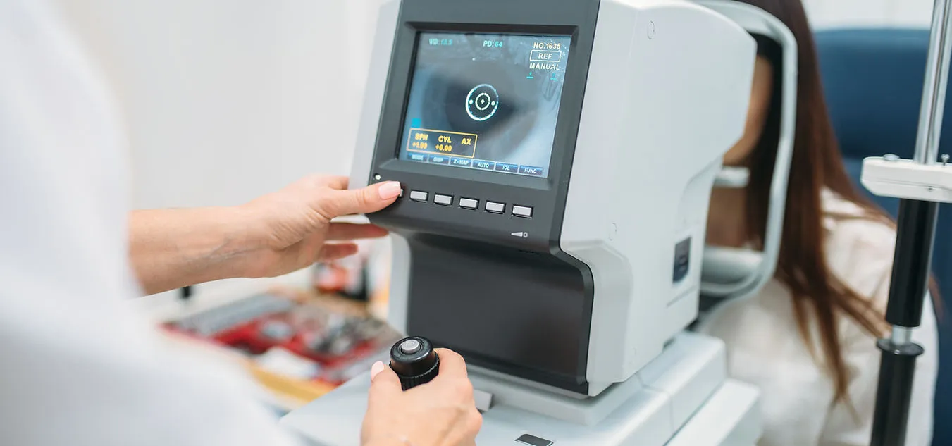
Our eye imaging technology
Every eye examination includes OCT scans and Optomap imaging.
Know your eye health
Traditional retinal imaging typically provides your optometrist with a 45° view of the retina – though this can be more restricted with smaller pupils. The optic nerve and the macula are the main details that are seen in these photos. By investing in OCT and Optomap technology that goes way beyond the traditional forms of capturing images, it allows us to detect and monitor any changes that may be indicative of a variety of pathologies, including glaucoma and macular degeneration

Best eyecare retinal imaging & Optomap imaging
The Optomap provides a much larger field of view of the retina, covering up to a 200° view of the retina.
This is generally not affected by pupil size so allows a thorough examination of the peripheral retina even with small pupils. In addition to allowing us to monitor and detect the same changes that can be seen with the conventional camera, Optomap’s wider field of view allows earlier detection of many systemic conditions such as high blood pressure, diabetes, high cholesterol levels, diabetic retinopathy and retinal tears/detachment.
For more information, click here to visit the Optomap website.
Best eyecare with enhanced imaging
OCT provides a radiation-free scan of your retina and creates a 3-dimensional image of the layers of your retina.
This allows conditions such as macular degeneration, diabetic retinopathy, glaucoma and vitreous detachment to be detected earlier and monitored with far greater accuracy than would be possible without it.